Pleural Mesothelioma Ct Images : Jcm Free Full Text When The Diagnosis Of Mesothelioma Challenges Textbooks And Guidelines Html / This scan also can show if it has spread to lymph nodes or other organs.
Pleural Mesothelioma Ct Images : Jcm Free Full Text When The Diagnosis Of Mesothelioma Challenges Textbooks And Guidelines Html / This scan also can show if it has spread to lymph nodes or other organs.. During stage 1 the tumor can also spread from the outer lining of the lung (closer to the wall of your chest) to the inner lining (closer to the lung itself). Early detection of the fatal and incurable mesothelioma and the subsequent provision of radiation, surgical and palliative asbestosis treatments are known to help a patient to have the best possible chance to extend and improve the quality of life remaining. Imaging is important in diagnosing mesothelioma.it will provide information such as the extent of disease in the original organ and also show if the cancer has spread to. To compare the multidetector ct (mdct) features of malignant pleural mesothelioma (mpm) and metastatic pleural disease (mpd). Than 1 cm, involvement of mediastinal pleural surface, are highly suggestive of malignant pleural disease, either mesothelioma or metastases.
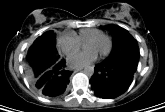
Doctors also use mesothelioma blood tests to live treatment response. Computed tomography of malignant pleural mesothelioma. It's the most common form of the cancer, comprising approximately 70 percent of diagnoses. This report describes the ct findings in five cases of pleural mesothelioma. Though these findings are frequently seen in mpm, they are not characteristic of mpm.
Mesothelioma Symptoms Diagnosis And Treatment Bmj Best Practice from bestpractice.bmj.com Because pleural mesothelioma occurs in several places in the lining of the lung simultaneously, surgery can be an ineffective treatment when used in isolation. Pseudomesotheliomatous lung cancer (plc), a term coined by harwood et al. A biopsy is the only definitive way to confirm a mesothelioma diagnosis. Or cat scans, and the more progressive pet/ct (positron emission tomography and computerized tomography). Analyzed multidetector ct images of 103 patients with malignant pleural mesothelioma and 24 patients with metastatic pleural disease from extrathoracic malignancy. 800cc of bloody, exudative fluid • pleural fluid cytology negative x 2 • thoracoscopy and right pleural biopsy performed: On ct a pleural plaque is defined as a discontinuous soft tissue focal thickening of the pleural surface, with or without foci of calcification. Imaging scans if an individual is experiencing mesothelioma cancer symptoms such as dysphagia, difficulty breathing, peritoneal or pleural effusion, chest pain, or wheezing, it is important to seek out a physical examination from a physician in order to properly diagnose these symptoms.additional techniques may be employed by the physician in order to correctly diagnose the problem.
From the study of the segmented bone several features can be extrapolated.
Jaklitsch mt, grondin sc, sugarbaker dj. A team of french and canadian researchers at laval university in quebec assessed the use of medical history and imaging features in patients with pleural thickening around the lungs. mesothelioma testing commonly includes imaging scans, biopsies and blood tests. Nakamori t, kosuda s, kyoto y, et al. Pet/ct scan challenge of pleural effusion treatment for mesothelioma patients. (b) ct scan image postcycle 2 of chemotherapy. J comput assist tomogr 1983; Although the chest film findings of pleural mesothelioma are well described, there are few descriptions of the findings of computed tomography (ct). From the study of the segmented bone several features can be extrapolated. pleural plaques occur when collagen, a protein, builds up in the lung lining. Ajr am j roentgenol 1981; A tissue biopsy will finalize whether or not a patient has this cancer. A new technique called ct perfusion can show if cancer cells are spreading in the bloodstream.
Using imaging tests to diagnose cancer is a field of medicine called diagnostic radiology. Analyzed multidetector ct images of 103 patients with malignant pleural mesothelioma and 24 patients with metastatic pleural disease from extrathoracic malignancy. Still, many doctors say the ct scan is the best for the chest and abdomen, which are where mesothelioma forms. mesothelioma testing commonly includes imaging scans, biopsies and blood tests. pleural mesothelioma is caused by inhaling asbestos fibers that get lodged into the protective lining of the lungs (the pleura) which cause genetic mutations in the surrounding cells.
Malignant Pleural Mesothelioma Radiology Case Radiopaedia Org from prod-images-static.radiopaedia.org Or cat scans, and the more progressive pet/ct (positron emission tomography and computerized tomography). images hosted on other servers: J comput assist tomogr 1983; Ipsilateral pleural effusion/ hydropneumothorax • ct chest: 30 patients with digitally available chest computed tomography (ct) scans before and after three cycles of chemotherapy were included. It's the most common form of the cancer, comprising approximately 70 percent of diagnoses. The segmentation of the rib cage in ct images represents a task of primary importance in medical imaging for different reasons. These features are indices of the presence of some diseases such as the malignant pleural mesothelioma (mpm) which is the main focus of our research.
Pet/ct examine challenge of pleural effusion treatment for mesothelioma patients. Malignant pleural mesothelioma is a rare tumor. Early detection of the fatal and incurable mesothelioma and the subsequent provision of radiation, surgical and palliative asbestosis treatments are known to help a patient to have the best possible chance to extend and improve the quality of life remaining. The ct and mri features of malignant mesothelioma are similar to those seen with other causes of malignant pleural thickening. While looking at the images from all scans and tests, mesothelioma specialists will rule out the possibility of lung cancer if there is a history of asbestos exposure. Pet/ct scan challenge of pleural effusion treatment for mesothelioma patients. Ajr am j roentgenol 1981; Ipsilateral pleural effusion/ hydropneumothorax • ct chest: These features are indices of the presence of some diseases such as the malignant pleural mesothelioma (mpm) which is the main focus of our research. It's in the lining of only one side of. Complementary to contrast enhanced ct. ct scans can locate pleural disease, chest wall invasions, and can also be used to guide fine needle aspiration tests. Doctors also use mesothelioma blood tests to measure treatment response.
800cc of bloody, exudative fluid • pleural fluid cytology negative x 2 • thoracoscopy and right pleural biopsy performed: Doctors also use mesothelioma blood tests to live treatment response. Mirvis s, dutcher jp, haney pj, et al. mesothelioma testing commonly includes imaging scans, biopsies and blood tests. The segmentation of the rib cage in ct images represents a task of primary importance in medical imaging for different reasons.
Clinical Staging Of Malignant Pleural Mesothelioma Current Perspectiv Lctt from www.dovepress.com The segmentation of the rib cage in ct images represents a task of primary importance in medical imaging for different reasons. pleural plaques occur when collagen, a protein, builds up in the lung lining. Only pleural mesothelioma, the kind in your lungs, has stages. Diagnosing pleural plaques simple exposure to asbestos does not mean that mesothelioma or other serious diseases should be the assumed diagnosis. The ct and mri features of malignant mesothelioma are similar to those seen with other causes of malignant pleural thickening. Doctors also use mesothelioma blood tests to live treatment response. Although the chest film findings of pleural mesothelioma are well described, there are few descriptions of the findings of computed tomography (ct). It's in the lining of only one side of.
These imaging tests will show visual evidence of mesothelioma.
Early detection of the fatal and incurable mesothelioma and the subsequent provision of radiation, surgical and palliative asbestosis treatments are known to help a patient to have the best possible chance to extend and improve the quality of life remaining. images hosted on other servers: The united kingdom has the highest rate with 8.2 pleural patients per 100,000 people in the country. In each case the ct showed. Jaklitsch mt, grondin sc, sugarbaker dj. pleural plaque is a harmless disease caused by asbestos exposure. pleural thickening/effusion can be distinguished with ct scanning.nodular pleural thickening, thickening > Grant dc, seltzer se, antman kh, et al. The ct and mri features of malignant mesothelioma are similar to those seen with other causes of malignant pleural thickening. pleural plaques occur when collagen, a protein, builds up in the lung lining. Because pleural mesothelioma is commonly misdiagnosed at first, doctors now use many imaging tests to accurately identify and stage mesothelioma. Doctors also use mesothelioma blood tests to live treatment response. Malignant pleural mesothelioma (mpm) is the most common type and can be difficult to treat because most patients have advanced disease at presentation.


0 Comments